Organoids made from amniotic fluid will tell us how fetuses develop
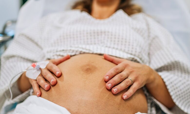
Researchers have known for decades that amniotic fluid holds fetal cells. That’s what allows doctors to diagnose conditions like Down syndrome and sickle-cell disease before birth via amniocentesis, in which a needle is used to take a sample of the fluid. The vast majority of these cells, 95% or more, are dead cells sloughed off by the fetus, says Mattia Gerli, a stem cell biologist at University College London and an author of a paper on the work published in Nature Medicine today. But what the researchers homed in on was the much smaller fraction of live cells in amniotic fluid.
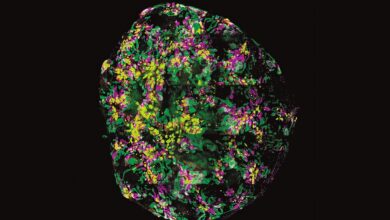
First, they worked to determine what kinds of cells were there, mapping their identities and then using single-cell sequencing to assess where they originated. Next, the team placed three kinds of progenitor cells—kidney, lung, and small intestine—in a 3D culture to see if they would form organoids.
“We’re just taking them as they are and putting them into a droplet of gel. This is very low tech,” coauthor Paolo De Coppi, a pediatric surgeon at University College London and the Great Ormond Street Hospital, said in a press briefing.
It worked. The organoids grew, and they developed features of the tissue that the cells came from. Within weeks the lung organoids, for example, had beating, hairlike structures called cilia, like those found inside the lung.